
The pupillary light reflex is used to evaluate brainstem functions in patients with suspected traumatic brain injuries (TBIs). This examination assists medical professionals in diagnosing TBIs and monitoring their progress. In this article, we will discuss the role of the pupillary light reflex in diagnosing TBIs.
By understanding how this test works, medical professionals can accurately assess whether a patient has suffered from head trauma and provide appropriate treatment options depending on the pupil measurement results. Furthermore, they can also monitor progress over time using repeated assessments.
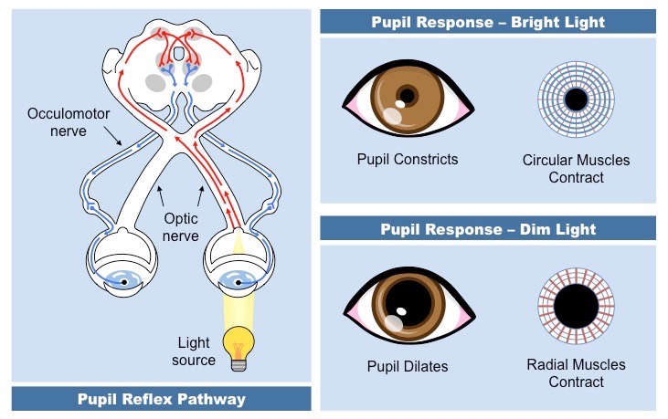
What Is the Pupillary Light Reflex?
The pupillary light reflex is remarkable, illuminating the intricate connection between our eyes and brains. Neuro exam results are often used to detect changes in this crucial pathway that can indicate various neurological disorders.
This test assesses the response of one’s pupils when exposed to bright light, allowing for an accurate diagnosis of certain conditions. It involves measuring pupil size and the differences between each eye’s response, thus providing information about neurological activity.
In addition, it may also reveal ocular mobility issues, which can further inform diagnostic decisions. The pupillary light reflex is integral in helping medical professionals diagnose TBIS because it provides detailed insight into neurological functioning.
How Is the Pupillary Light Reflex Used to Diagnose TBIs?
The pupillary light reflex is an important diagnostic tool for medical professionals. It utilizes the size of pupils in response to a bright light as an indication of neurological integrity and potential signs of disease or trauma. For example, suppose a patient was recently diagnosed with a traumatic brain injury (TBI).
Upon examination of the pupillary response in traumatic brain injury, the physician would observe that when presented with a bright light, one pupil responded significantly slower than the other, indicating possible damage to her nervous system due to TBI. This observation led to further tests, which revealed significant tissue damage in certain areas of her brain.
This reflex has also been used effectively to diagnose diseases such as Parkinson’s and Multiple Sclerosis, which can cause changes in pupil responses to light stimuli. In addition, it has been noted that patients suffering from migraine headaches often display abnormal pupillary constriction and dilatation patterns during these episodes.
In sum, the pupillary light reflex and other neurological tools, such as the NPi pupilometer are invaluable for diagnosing various neurological disorders and diseases and determining levels of intoxication through their ability to measure minute differences in pupil sizes when exposed to different lighting conditions. Moving forward, we must explore the significance of this reflex in diagnosing TBIs specifically.
The Significance of Pupillary Light Reflex in Diagnosing TBIs
The pupillary light reflex has been a reliable diagnostic tool since the early 1800s. It measures pupil size in response to light, revealing signs of neurological damage or compression on the optic nerve. In addition, changes in pupil size can be indicators of TBI, MS, or Parkinson’s. With a penlight and exam room lighting, clinicians can access crucial information about a patient’s health. Thus, this ancient technique should not be overlooked; its value lies in providing better care and outcomes for those with neurological issues.
The Limitations of Pupillary Light Reflex in Diagnosing TBIs
Pupillary light reflex has long been used as a diagnostic tool in the medical field, but there are limitations to its effectiveness. Despite its utility and accuracy in determining neurological conditions, the pupillary light reflex has limitations that may affect how accurately doctors can diagnose TBIs.
One limitation of this test is that pupil diameter does not necessarily reveal the underlying cause of an issue; for example, pupil constriction due to bright lights will show up regardless of whether or not there is a neurological disorder present. Additionally, since pupils naturally fluctuate in size depending on ambient lighting levels and other factors, it can be difficult to discern any changes caused by a neurological condition from normal variability.
Finally, pupil responses depend on patient cooperation; if someone fails to follow instructions properly, the pupillary light reflex test results may be inaccurate.
Doctors should consider these limitations when utilizing the pupillary light reflex to diagnose TBIs. As such, practitioners must determine if other alternatives provide more accurate information when assessing cases of suspected TBI.
- How to Prepare for a Cyber Security Job Interview - June 15, 2023
- Unblocked Games: Unlocking Fun and Learning Without Restrictions - June 14, 2023
- The 10 Principles of Insider Risk Management - June 14, 2023